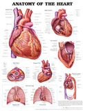
40 the human heart and its labels
Conceptual metaphor - Wikipedia In cognitive linguistics, conceptual metaphor, or cognitive metaphor, refers to the understanding of one idea, or conceptual domain, in terms of another.An example of this is the understanding of quantity in terms of directionality (e.g. "the price of peace is rising") or the understanding of time in terms of money (e.g."I spent time at work today"). Human Heart - Anatomy, Functions and Facts about Heart The human heart is one of the most important organs responsible for sustaining life. It is a muscular organ with four chambers. The size of the heart is the size of about a clenched fist. The human heart functions throughout a person’s lifespan and is one of the most robust and hardest working muscles in the human body.
Human Heart Diagram - Human Body Pictures - Science for Kids Find free pictures, photos, diagrams, images and information related to the human body right here at Science Kids. Photo name: Human Heart Diagram Picture category: Human Body Image size: 70 KB Dimensions: 600 x 600 Photo description: This is an excellent human heart diagram which uses different colors to show different parts and also labels a number of important heart component such as the ...

The human heart and its labels
Heart Diagram with Labels and Detailed Explanation The human heart is the most crucial organ of the human body. It pumps blood from the heart to different parts of the body and back to the heart. The most common heart attack symptoms or warning signs are chest pain, breathlessness, nausea, sweating etc. The diagram of heart is beneficial for Class 10 and 12 and is frequently asked in the ... Labelling the heart — Science Learning Hub Blood transports oxygen and nutrients to the body. It is also involved in the removal of metabolic wastes. In this interactive, you can label parts of the human heart. Drag and drop the text labels onto the boxes next to the diagram. Selecting or hovering over a box will highlight each area in the diagram. How to Draw a Human Heart: 11 Steps (with Pictures) - wikiHow If you're trying to identify parts of the heart for a class you're taking, it's good practice to draw the heart yourself and label each segment. You can refer to your textbook in order to label the: Aorta Superior vena cava Inferior vena cava Right and left atria Right and left ventricles Pulmonary veins and arteries 5
The human heart and its labels. Oracle Human Resources Modules Our complete human capital management (HCM) solution in the cloud is an intelligent, personalized way to adapt faster. At the core of our HCM solution is Oracle Human Resources. This innovative family of product capabilities uses AI and machine learning to help you plan, manage, and optimize HR practices for a global and diverse workforce all in a single system. It … Anatomy of the Human Heart - Physiopedia The heart is a muscular organ that serves to collect deoxygenated blood from all parts of the body, carries it to the lungs to be oxygenated and release carbon dioxide. Then, it transports the oxygenated blood from the lungs and distributes it to all the body parts. The heart pumps around 7,200 litres of blood in a day throughout the body.; The heart is situated at the centre of the chest and ... Heart: Anatomy and Function Your heart is the main organ of your cardiovascular system, a network of blood vessels that pumps blood throughout your body. It also works with other body systems to control your heart rate and blood pressure. Your family history, personal health history and lifestyle all affect how well your heart works. Appointments 800.659.7822 commons.wikimedia.org › wiki › File:Diagram_of_theFile:Diagram of the human heart (cropped).svg - Wikimedia Apr 05, 2022 · English: Diagram of the human heart 1. Superior vena cava 2. 4. Mitral valve 5. Aortic valve 6. Left ventricle 7. Right ventricle 8. Left atrium 9. Right atrium 10. Aorta 11. Pulmonary v
Diagram of the human heart Images, Stock Photos & Vectors ... 14,495 diagram of the human heart stock photos, vectors, and illustrations are available royalty-free. See diagram of the human heart stock video clips Image type Orientation Sort by Popular Anatomy Healthcare and Medical Diseases, Viruses, and Disorders Icons and Graphics heart medicine organ human body circulatory system diagram Next of 145 & Stroke Foundation of Barbados Inc.: Everybody Has A ... On April 18, 2020, the Heart and Stroke Foundation of Barbados Inc. celebrated 35 years of endorsing healthy lifestyles and hearts! Our journey began in 1985, and to date we have supported thousands of Barbadians by providing training, education campaigns and free or subsidized services to aid in combating heart disease. Draw And Label The Human Heart - Draw And Label External ... Diagram of the human heart. How can i draw a diagram of the human heart and label its different parts? Click here to get an answer to your question ️ draw a diagram of the human heart and label its parts. It is situated between the lungs in the upper thoracic cavity at the . You probably have a lot of experience drawing a cartoon heart with the. Human Genome Project Results 12.11.2018 · The finished sequence produced by the Human Genome Project covers about 99 percent of the human genome's gene-containing regions, and it has been sequenced to an accuracy of 99.99 percent. In addition, to help researchers better understand the meaning of the human genetic instruction book, the project took on a wide range of other goals, from …
PDF Anatomy of Heart Labeled and Unlabeled Images aortic arch left pulmonary artery left pulmonary veins auricle of left atrium left atrium circumflex artery (in atrioventricular sulcus) coronary sinus left ventricle (c) posterior view of the external heart © 2019 pearson education, inc. ascending aorta superior vena cava right pulmonary artery right pulmonary veins right atrium inferior vena … Human heart: Anatomy, function & facts | Live Science The human heart is an organ that pumps blood throughout the body via the vessels of the circulatory system, supplying oxygen and nutrients to the tissues and removing carbon dioxide and other ... en.wikipedia.org › wiki › Human_eyeHuman eye - Wikipedia Each eye has seven extraocular muscles located in its orbit. Six of these muscles control the eye movements, the seventh controls the movement of the upper eyelid.The six muscles are four recti muscles – the lateral rectus, the medial rectus, the inferior rectus, and the superior rectus, and two oblique muscles the inferior oblique, and the superior oblique. Anatomy of a Human Heart - uofmhealth Located between the lungs in the middle of the chest, the heart pumps blood through the network of arteries and veins known as the cardiovascular system. It pushes blood to the body's organs, tissues and cells. Blood delivers oxygen and nutrients to every cell and removes the carbon dioxide and other waste products made by those cells.

Congestive Heart Failure: The Essence of Heart Failure Course | CEUfast Nursing Continuing Education
File:Diagram of the human heart (no labels).svg ... File:Diagram of the human heart (no labels).svg. Size of this PNG preview of this SVG file: 498 × 599 pixels. Other resolutions: 199 × 240 pixels | 399 × 480 pixels | 499 × 600 pixels | 639 × 768 pixels | 851 × 1,024 pixels | 1,703 × 2,048 pixels | 533 × 641 pixels.
Normal Anatomy of the Human Heart Giclee Print by Nucleus Medical Art - at AllPosters.com.au
Heart: illustrated anatomy - e-Anatomy 11 - Proximal circumflex 11. Mid inferolateral 12 - Intermediate/anterolateral 12. Mid anterolateral 12a - Obtuse marginal a 12b - Obtuse marginal b 13 - Distal circumflex 13. Apical anterior 14 - Left posterolateral 14. Apical septal 14a - Left posterolateral a 14b - Left posterolateral b 15 - Posterior descending 15. Apical inferior
Sampoerna Wallpaper: The Heart Diagram Labeled
(PDF) Human Dignity and Human Rights - ResearchGate Specifically what I want to show is: 1. that the idea of human dignity plays a decisive role in today’s social and political thought and action, even more so than commonly realized; 2. that this ...
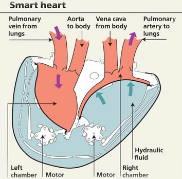
Our World | What is the heart?
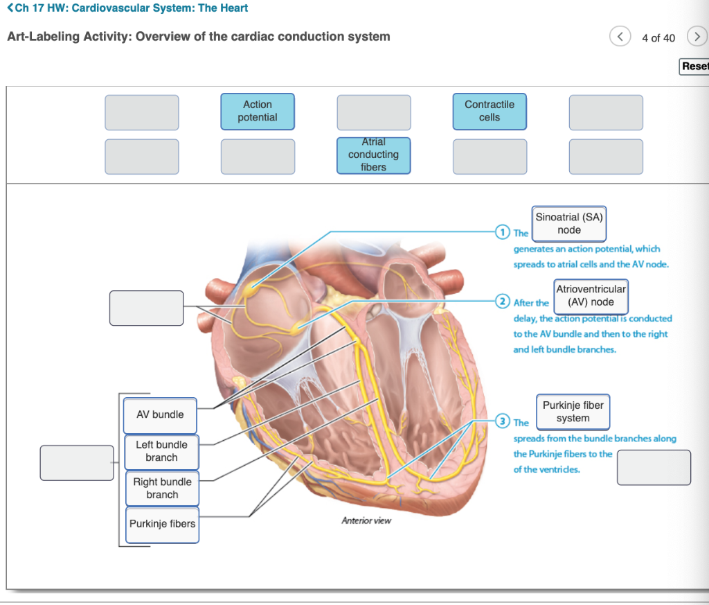
Heart - Wikipedia The wall of the heart is made up of three layers: epicardium, myocardium, and endocardium. The heart pumps blood with a rhythm determined by a group of pacemaker cells in the sinoatrial node. These generate a current that causes the heart to contract, traveling through the atrioventricular node and along the conduction system of the heart.

Parts Of The Human Heart | Science Trends
› health › educationalAssessing Your Weight and Health Risk It is calculated from your height and weight. BMI is an estimate of body fat and a good gauge of your risk for diseases that can occur with more body fat. The higher your BMI, the higher your risk for certain diseases such as heart disease, high blood pressure, type 2 diabetes, gallstones, breathing problems, and certain cancers.

Human Heart Diagram Educational Posters EP126
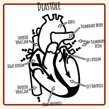
PDF Analyzing the Human Heart - Beyond the Classroom - Home working. Its job is to pump blood to the lungs and to all of the body tissues. In this activity you will use a diagram of the heart to analyze the way in which the heart works. l. Using the following word list, label the various parts of the heart on the diagram. Right ventricle Left venfficle Upper vena cava Lower vena cava Aorta

Simplified Heart Labeled Decal | Shop Fathead Anatomical Images Graphics
Heart Diagram - 15+ Free Printable Word, Excel, EPS, PSD ... Teachers and students use the heart diagram, in biological science, to study the structure and functions of a human being's heart. Friends and colleagues on the other hand may find this diagram template useful when it comes to sending special, personalized gifts to their family members and significant others.

Human Nervous System Structure and Functions Explained With Diagrams - Bodytomy
Heart histology: Cells and layers | Kenhub The heart is a critical organ that keeps blood moving throughout the body. Blood is an important medium that not only carries nutrients and oxygen throughout the body, but it also collects waste products and returns them to the liver and kidney for further processing and excretion.. The heart is able to achieve this autonomy based on its histological make-up.

Anatomy and Physiology e-Portfolio: December 2010
The Anatomy of the Heart, Its Structures, and Functions The heart is the organ that helps supply blood and oxygen to all parts of the body. It is divided by a partition (or septum) into two halves. The halves are, in turn, divided into four chambers. The heart is situated within the chest cavity and surrounded by a fluid-filled sac called the pericardium. This amazing muscle produces electrical ...
:max_bytes(150000):strip_icc()/human-heart-circulatory-system-598167278-5c48d4d2c9e77c0001a577d4.jpg)
The Anatomy of the Heart, Its Structures, and Functions
Human Heart - Diagram and Anatomy of the Heart The heart is a muscular organ about the size of a closed fist that functions as the body's circulatory pump. It takes in deoxygenated blood through the veins and delivers it to the lungs for oxygenation before pumping it into the various arteries (which provide oxygen and nutrients to body tissues by transporting the blood throughout the body).
Human Heart - Label the Diagram Anatomy Clip Art Set Commercial Use
Human Heart Diagram Labeled | Science Trends Human Heart Diagram Labeled Daniel Nelson 1, January 2019 | Last Updated: 3, March 2020 The human heart is an organ responsible for pumping blood through the body, moving the blood (which carries valuable oxygen) to all the tissues in the body. Without the heart, the tissues couldn't get the oxygen they need and would die.

Parts Of Heart Diagram Stock Illustration - Download Image Now - iStock
Diagram of Human Heart and Blood Circulation in It | New ... Exterior of the Human Heart A heart diagram labeled will provide plenty of information about the structure of your heart, including the wall of your heart. The wall of the heart has three different layers, such as the Myocardium, the Epicardium, and the Endocardium. Here's more about these three layers. Epicardium

New Photos in Anatomy of human body organs
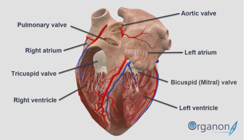
A Labeled Diagram of the Human Heart You Really Need to ... The human heart, comprises four chambers: right atrium, left atrium, right ventricle and left ventricle. The two upper chambers are called the left and the right atria, and the two lower chambers are known as the left and the right ventricles. The two atria and ventricles are separated from each other by a muscle wall called 'septum'.

Human Heart Pictures with Labels Best Of File Diagram Of the Human Heart Hug Wikimedia Mons ...
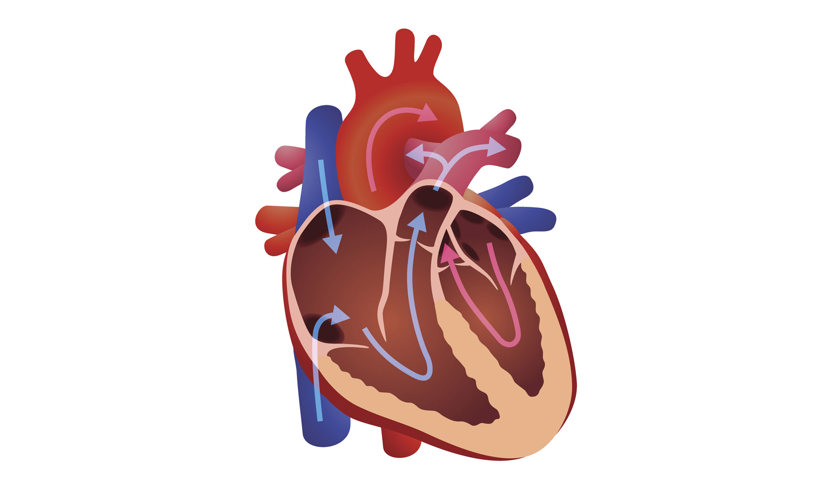
A Diagram of the Heart and Its Functioning Explained in ... The heart blood flow diagram (flowchart) given below will help you to understand the pathway of blood through the heart.Initial five points denotes impure or deoxygenated blood and the last five points denotes pure or oxygenated blood. 1.Different Parts of the Body ↓ 2.Major Veins ↓ 3.Right Atrium ↓ 4.Right Ventricle ↓ 5.Pulmonary Artery ↓ 6.Lungs
How the human heart did become associated with love? | 3D ORGANON
news.yahoo.com › fda-identifies-2-red-flagFDA identifies 2 red-flag words to look out for on your ... Apr 25, 2022 · The FDA found Ortiga products also typically contain diclofenac, which can increase the risk of cardiovascular events like stroke and heart attack, and may lead to ulceration or fatal perforation of the stomach and intestines. Dexamethasone, a corticosteroid that has been used to treat severely ill COVID-19 patients on ventilators. It can ...
32 Label This Anterior View Of The Human Heart - Labels Design Ideas 2020
Label the heart — Science Learning Hub Label the heart Add to collection In this interactive, you can label parts of the human heart. Drag and drop the text labels onto the boxes next to the diagram. Selecting or hovering over a box will highlight each area in the diagram. Pulmonary vein Right atrium Semilunar valve Left ventricle Vena cava Right ventricle Pulmonary artery Aorta

Pokemon Cosplay: May 2014
Human Heart (Anatomy): Diagram, Function, Chambers ... The heart is a muscular organ about the size of a fist, located just behind and slightly left of the breastbone. The heart pumps blood through the network of arteries and veins called the...
Post a Comment for "40 the human heart and its labels"