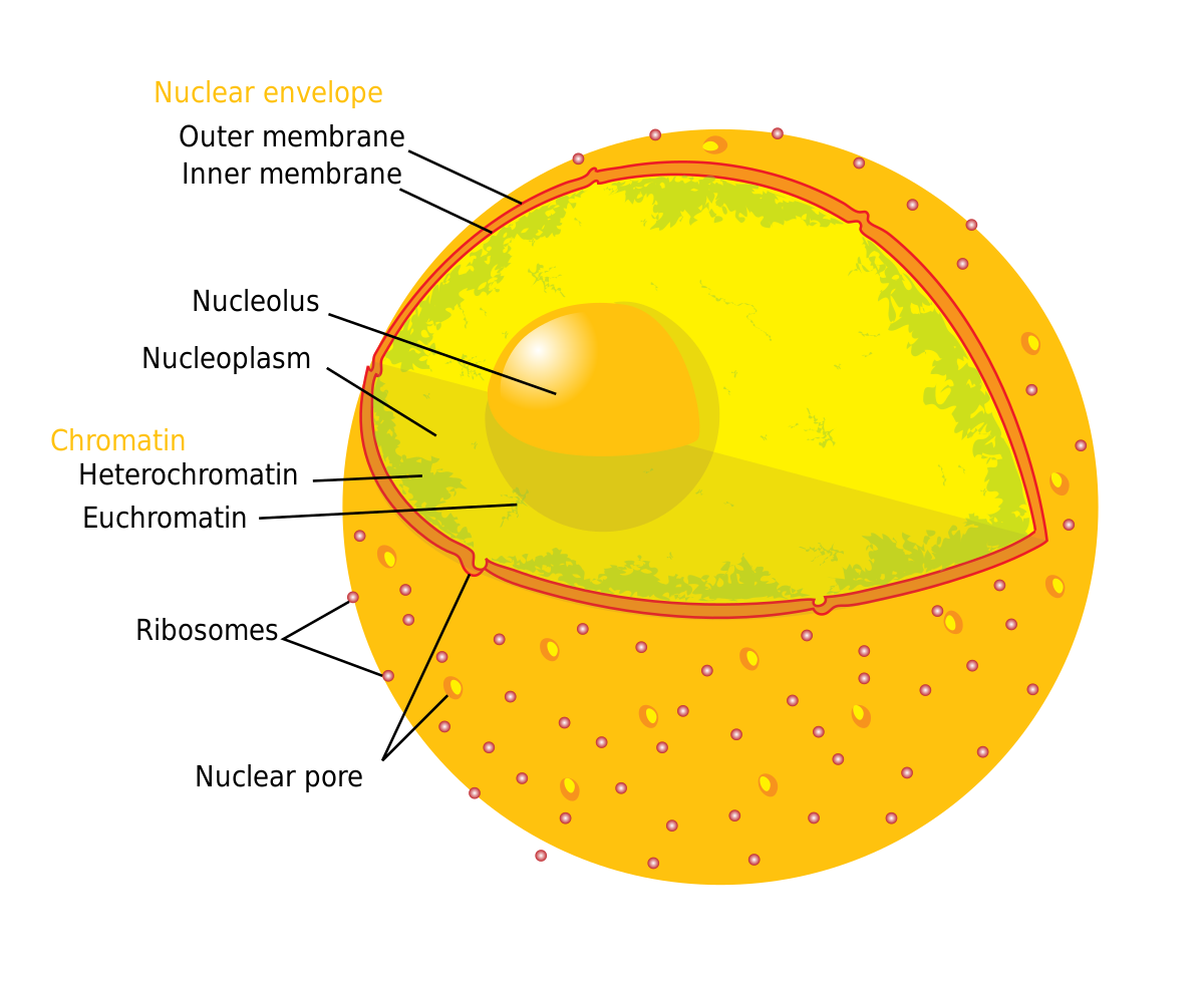
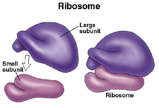
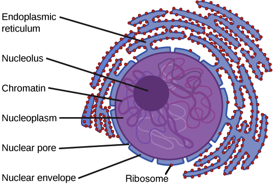
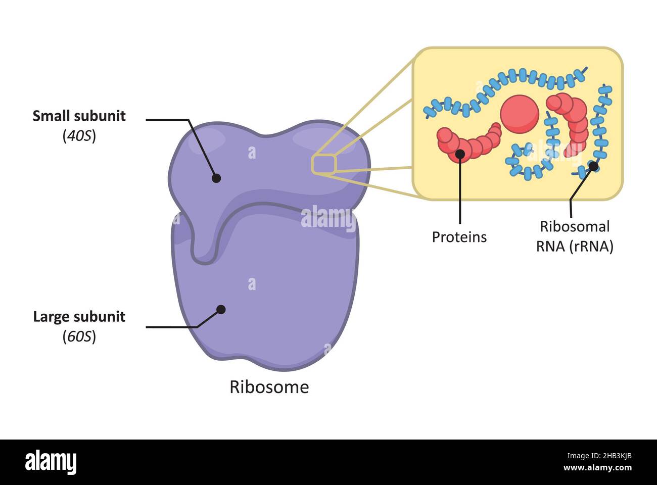
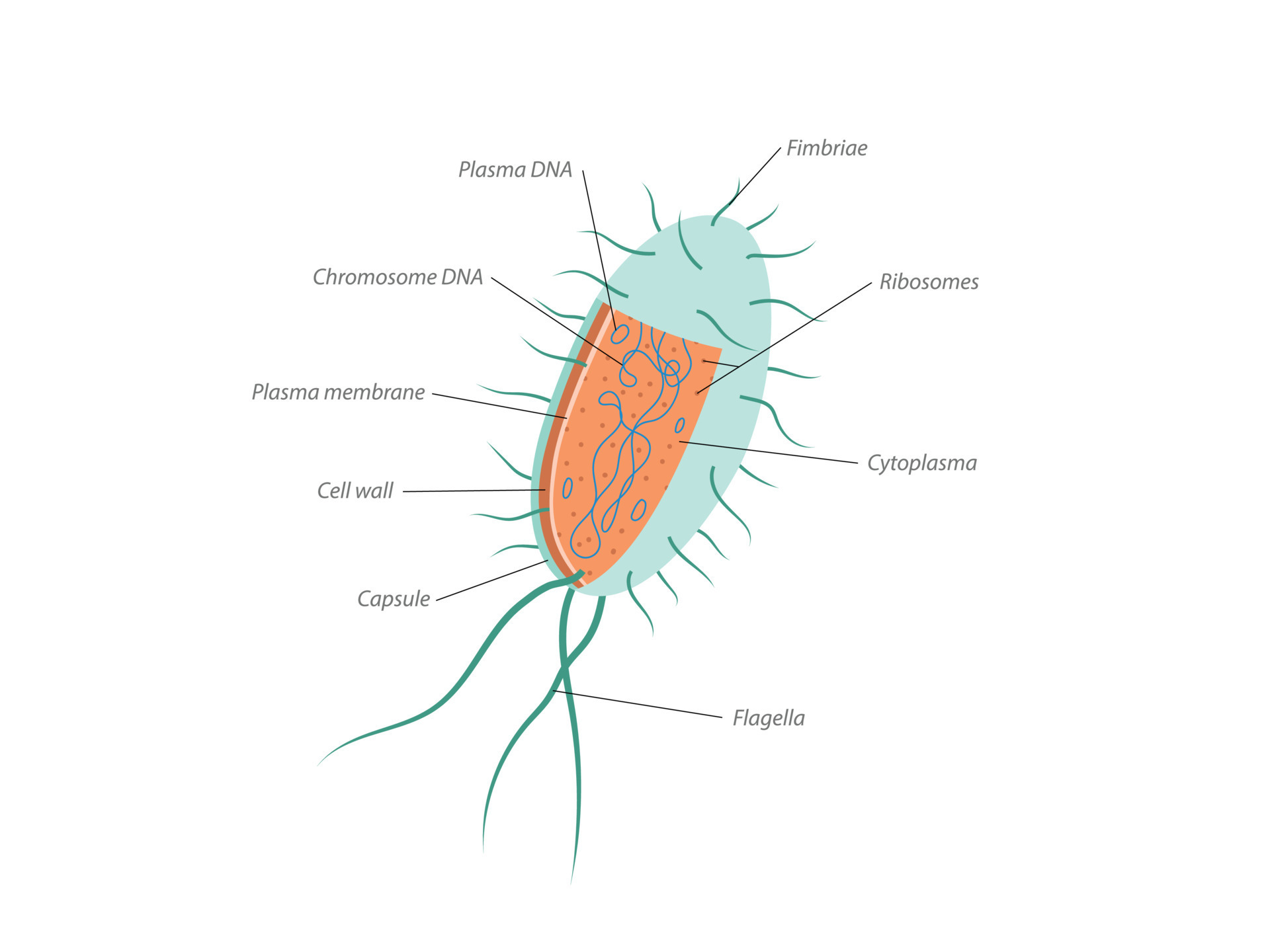
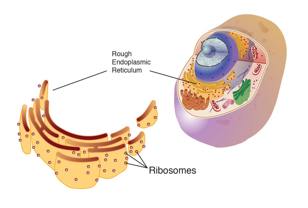
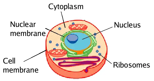
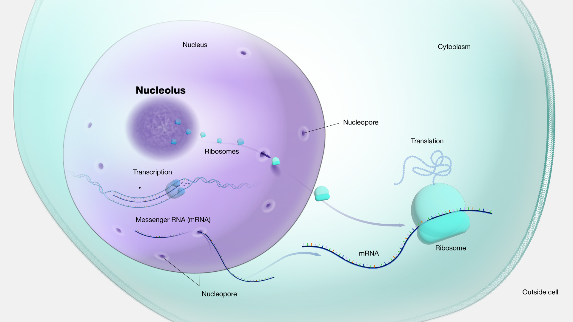
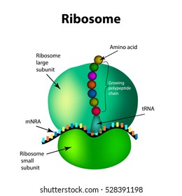
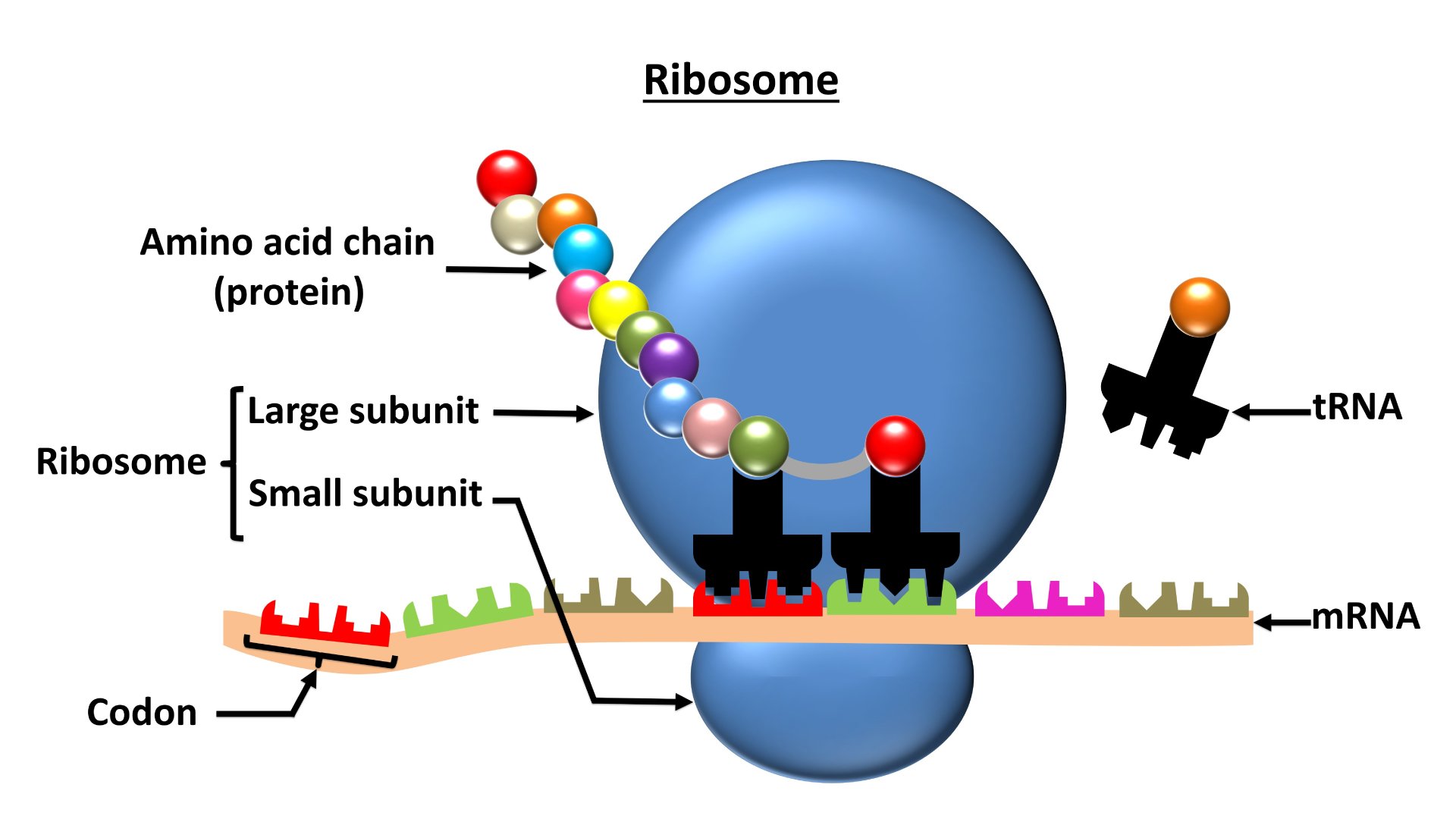
40 ribosome diagram with labels
Biology 1005 - Chapter 6 DNA: The Molecule of Life - Quizlet Study with Quizlet and memorize flashcards containing terms like Can you complete this paragraph about the cellular processes involved in protein production?, The cellular processes that results in the production of protein begin in the _____ , where the DNA resides. There, the process of _____ creates a molecule of RNA from a molecule of DNA. The enzyme that performs this process is called ... Bio101 - Ch 6 HW Flashcards | Quizlet Study with Quizlet and memorize flashcards containing terms like Which of the following choices correctly matches a tool and its proper application? See Concept 6.1 -cell fractionation to study the function of specific organelles -light microscopy to study the internal structure of cilia -transmission electron microscopy (TEM) to study the surfaces of preserved cells -transmission electron ...
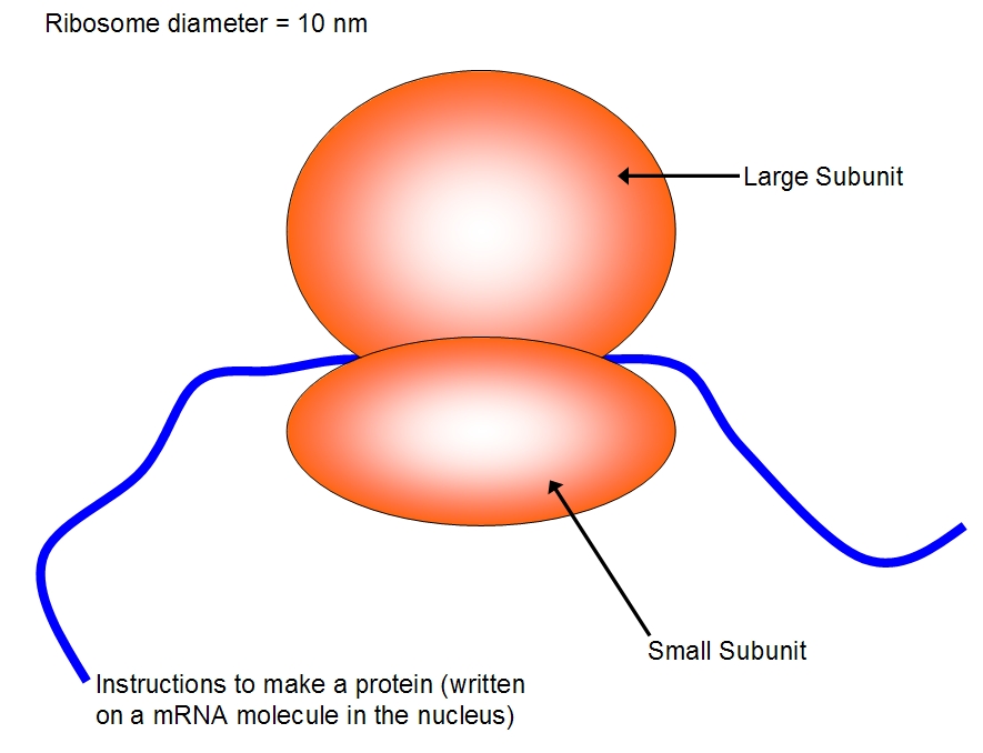
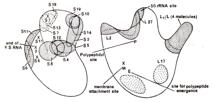
Structure of Ribosome (With Diagram) - Biology Discussion A bacterial ribosome is about 250 nm in diameter and consists of two subunits, one large and one small. Both subunits consist of one or more molecules of rRNA and an array of ribosomal proteins. ADVERTISEMENTS: Association of two subunits is called mono-some. The structure of prokaryotic ribosome is given in the figure 8.2 B.
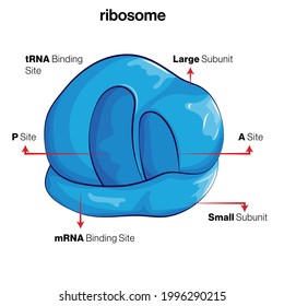
Ribosome diagram with labels
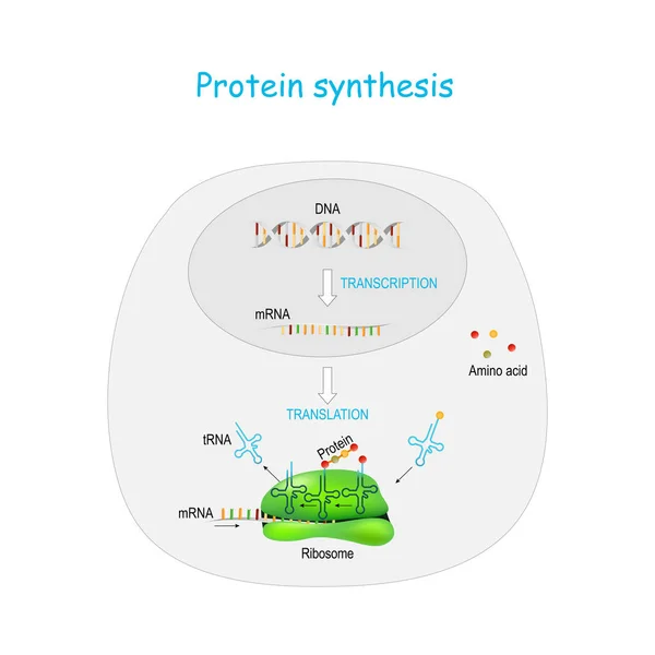
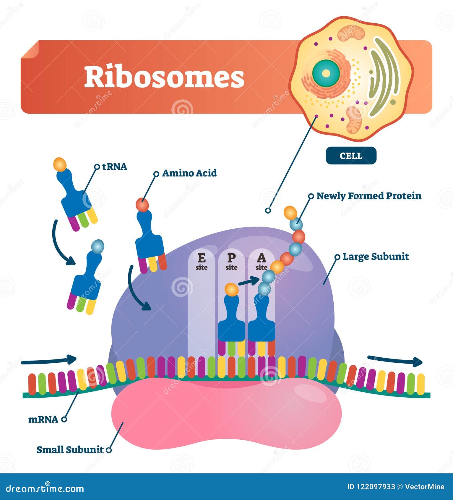
Ribosome and protein synthesis, diagram - Stock Image - C029/3020 Messenger ribonucleic acid (mRNA, blue with coloured nucleotides) is read by a ribosome (pink). The molecules of transfer RNA (tRNA, key-shaped) each bring an amino acid (orange dot) to bind to the ribosome's protein synthesis site. The amino acid added is determined by the three-nucleotide coding (codon) on the mRNA, which matches up with the ... Bio 1113 - Unit 11 - Gene Expression Flashcards | Quizlet In the following diagram of a ribosome, assign the correct labels: Label 1: a tRNA attached to a polypeptide is found in this area of the ribosome Label 2: a tRNA attached to a single amino acid enters here Label 3: a tRNA that is not attached to anything exits here Label 4: a tRNA molecule Label 5: growing polypeptide Label 6: mRNA being ... What Are Ribosomes? - Definition, Structure and its Functions - BYJUS Ribosomes are located inside the cytosol found in the plant cell and animal cells. The ribosome structure includes the following: It is located in two areas of cytoplasm. Scattered in the cytoplasm. Prokaryotes have 70S ribosomes while eukaryotes have 80S ribosomes. Around 62% of ribosomes are comprised of RNA, while the rest is proteins.
Ribosome diagram with labels. Mastering Biology Chapter 6 Flashcards | Quizlet Study with Quizlet and memorize flashcards containing terms like Which of the following choices correctly matches a tool and its proper application? a. light microscopy to study the internal structure of cilia b. scanning electron microscopy (SEM) to study the detailed movements of living cells c. transmission electron microscopy (TEM) to study the surfaces of preserved cells d. cell ... BIONIC: biological network integration using convolutions ... Oct 03, 2022 · A Venn diagram describes the overlap of captured complexes (score of 0.5 or higher) between the input networks and BIONIC integration. ... When gene labels are provided, an additional ... Exam 3 Flashcards | Quizlet The ribosome in the diagram is in the process of synthesizing a protein using directions transcribed from the DNA. Use the labels to identify each of the structures involved in translation and protein synthesis. Ribosomes: Structure, Composition, and Assembly (With Diagram) Ribosomes: Structure, Composition, and Assembly (With Diagram) Article Shared by ADVERTISEMENTS: We will be concerned with the organization, composition, and assembly of the cytoplasmic ribosomes of prokaryotic and eukaryotic cells. The ribosomes present in cytoplasmic organelles (e.g., chloroplast and mitochondrial ribosomes).
Molecular Modeling Database (MMDB) Help Document Example: The ribosome structure by Selmer, Dunham, Murphy, Weixlbaumer, Petry, Kelley, Weir, and Ramakrishnan, the 2009 Nobel Laureate in Chemistry, was split into PDB records 2XFZ, 2XG0, 2XG1, 2XG2, and was merged at MMDB into a single record with the MMDB ID 99580: Mastering Quiz: Chapter 7A Microbial Genetics Flashcards | Quizlet Those segments of the RNA strand that do not actually code for the protein are removed. c. mRNA binds to a ribosome in the cytoplasm. d. A molecule of RNA is formed based on the sequence of nucleotides in DNA. e. ... Drag the correct labels under the diagrams to identify the events of RNA processing. Drag the labels onto the diagram to identify ... Solved In the following diagram of a ribosome, assign the - Chegg Question: In the following diagram of a ribosome, assign the correct labels. ... In the following diagram of a ribosome, assign the correct labels. Show transcribed image text Expert Answer. Who are the experts? Experts are tested by Chegg as specialists in their subject area. We review their content and use your feedback to keep the quality high. Solved The ribosome in the diagram is in the process of | Chegg.com Expert Answer 100% (264 ratings) Transcribed image text: The ribosome in the diagram is in the process of synthesizing a protein using directions transcribed from the DNA. Use the labels to identify each of the structures involved in translation and protein synthesis. Previous question Next question
Ribosome - Wikipedia Ribosomes consist of two major components: the small and large ribosomal subunits. Each subunit consists of one or more ribosomal RNA (rRNA) molecules and many ribosomal proteins (RPs or r-proteins). [1] [2] [3] The ribosomes and associated molecules are also known as the translational apparatus . Contents 1 Overview 2 Discovery 3 Structure Ribosome - protein factory - definition, function, structure and biology The protein translation by a ribosome consists of three stages: (1) Initiation, (2) Elongation, and (3) Termination. Initiation - the ribosome assembles around the target mRNA. A small ribosome subunit links onto the "start-end" of an mRNA strand. "Initiator tRNA" also enters the small subunit and binds to the start codon (most commonly, AUG). Structure of Ribosome - Biology Wise Ribosomes in Eukaryotes are 80S, where each one comprises 40S (small) and 60S (large) subunits. 60S comprises 5S subunit of RNA (almost 120 nucleotides) along with units of 28S RNA (almost 4700 nucleotides), subunit of 5.8S (close to 160 nucleotides) and ~49 units of protein. 40S has 18S RNA (approximately 1900 nucleotide) and ~33 units of protein. Ribosomes- Definition, Structure, Functions and Diagram - Microbe Notes Figure: Diagram of Ribosomes Structure of Ribosomes A ribosome is made from complexes of RNAs and proteins and is, therefore, a ribonucleoprotein. Around 37 to 62% of RNA is comprised of RNA and the rest is proteins. Each ribosome is divided into two subunits: A smaller subunit which binds to a larger subunit and the mRNA pattern, and
Ribosome Diagrams Ribosome Diagrams Ribosomes, Transcription, and Translation A two-part diagram shows both a complete ribosome (shown at right) and. Figure 7: The ribosome and translation. Ribosomes consist of two major components: the small and large ribosomal subunits. Each subunit consists of one or more ribosomal RNA (rRNA) molecules and many.
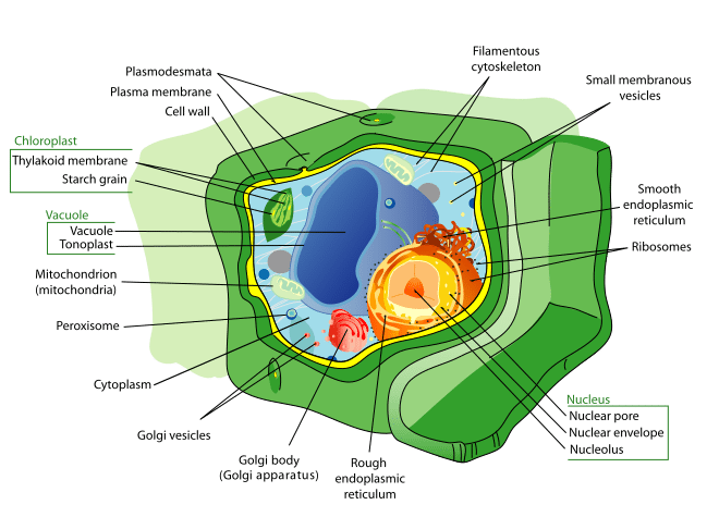
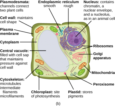
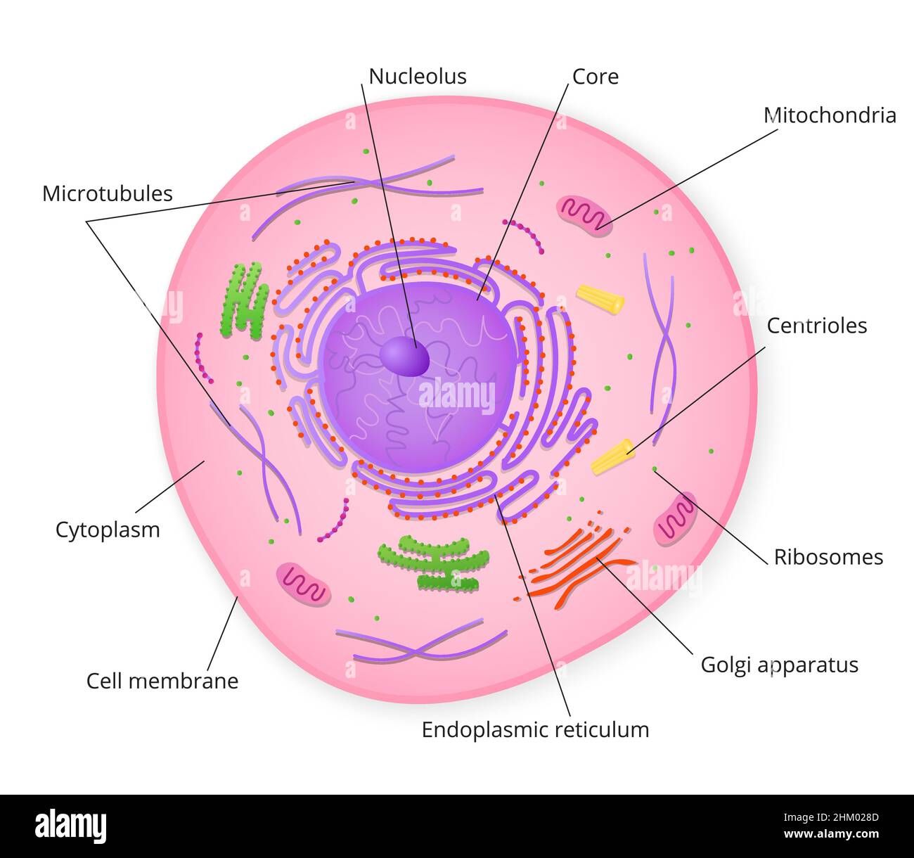
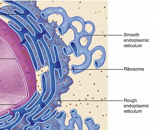
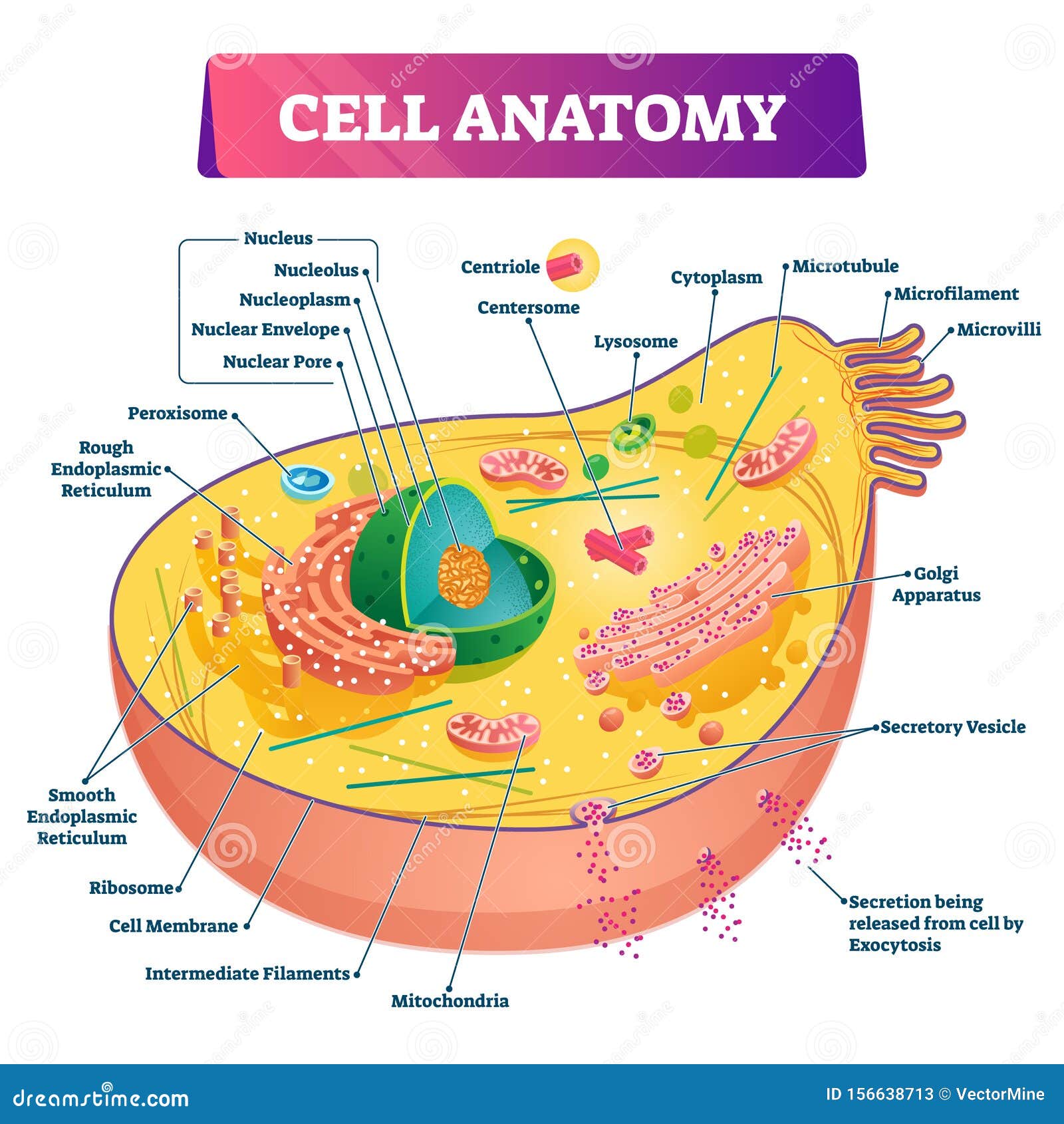
Animal Cell Ribosomes Diagram : Plant And Animal Cell Notes Notes ... Drag the labels on the left onto the diagram of the animal cell to correctly identify the function performed by each cellular structure. A bacterial ribosome is about 250 nm in diameter and consists of two subunits, one large …
Active Ribosome Profiling with RiboLace - PubMed Ribosome profiling, or Ribo-seq, is based on large-scale sequencing of RNA fragments protected from nuclease digestion by ribosomes. Thanks to its unique ability to provide positional information about ribosomes flowing along transcripts, this method can be used to shed light on mechanistic aspects …
The diagram at right represents a ribosome bound to The diagram at right represents a ribosome bound to mRNA a On the diagram label from LIFESCIENC 7A at University of California, Los Angeles. Study Resources. Main Menu; by School; by Literature Title; ... The diagram at right represents a ribosome bound to mRNA a On the diagram label.
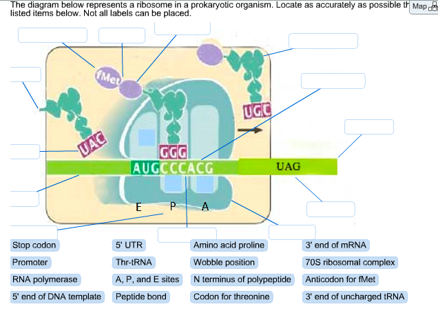
BIOL Chapter 12 Review Flashcards | Quizlet In the following diagram of a ribosome, assign the correct labels. A 5'-CUA-3' codon in an mRNA could be recognized by which of the following anticodon sequences in a tRNA? 3'-GAU-5' The reason that the genetic code can correctly specify the order of amino acids in a polypeptide is.
Protein Targeting (With Diagram) | Molecular Biology ADVERTISEMENTS: Let us make an in-depth study of the protein targeting. After reading this article you will learn about: 1. Introduction to Protein Targeting 2. Signal Sequence 3. Transport of Proteins into ER 4. Signal Sequence Recognition Mechanism 5. Role of Golgi Complex in Protein Transportation 6. Transport of Proteins from Golgi to Lysosomes 7. […]
What Are Ribosomes? - Definition, Structure and its Functions - BYJUS Ribosomes are located inside the cytosol found in the plant cell and animal cells. The ribosome structure includes the following: It is located in two areas of cytoplasm. Scattered in the cytoplasm. Prokaryotes have 70S ribosomes while eukaryotes have 80S ribosomes. Around 62% of ribosomes are comprised of RNA, while the rest is proteins.
Bio 1113 - Unit 11 - Gene Expression Flashcards | Quizlet In the following diagram of a ribosome, assign the correct labels: Label 1: a tRNA attached to a polypeptide is found in this area of the ribosome Label 2: a tRNA attached to a single amino acid enters here Label 3: a tRNA that is not attached to anything exits here Label 4: a tRNA molecule Label 5: growing polypeptide Label 6: mRNA being ...
Ribosome and protein synthesis, diagram - Stock Image - C029/3020 Messenger ribonucleic acid (mRNA, blue with coloured nucleotides) is read by a ribosome (pink). The molecules of transfer RNA (tRNA, key-shaped) each bring an amino acid (orange dot) to bind to the ribosome's protein synthesis site. The amino acid added is determined by the three-nucleotide coding (codon) on the mRNA, which matches up with the ...










Post a Comment for "40 ribosome diagram with labels"