40 microscope images with labels
Amazon.com: microscope slide labels Microscope Slide Label SLS-15, Standard, 1000/PK. $33.00 $ 33. 00. Get it Wed, Oct 19 - Mon, Oct 24. $8.00 shipping. Only 10 left in stock - order soon. Small Business. Small Business. Shop products from small business brands sold in Amazon's store. Discover more about the small businesses partnering with Amazon and Amazon's commitment to ... Microscope Labeling - The Biology Corner The google slides shown below have the same microscope image with the labels for students to copy. I often spend the first day walking students through the steps and having them look at a single slide as we do the steps. Students are often very enthusiastic about using microscopes and will try to start with the high power objective.
400+ Free Microscope & Bacteria Images - Pixabay 412 Free images of Microscope Related Images: bacteria laboratory science scientist research biology lab virus microscopic Find your perfect microscope image. Free pictures to download and use in your next project.
Microscope images with labels
Flow cytometry - Wikipedia Flow cytometry (FC) is a technique used to detect and measure physical and chemical characteristics of a population of cells or particles.. In this process, a sample containing cells or particles is suspended in a fluid and injected into the flow cytometer instrument. The sample is focused to ideally flow one cell at a time through a laser beam, where the light scattered is … Respiratory Histology Labeled | Virtual Anatomy Lab VAL - ncccval Body cavities, planes, and regions. Body Images Labeled. Body Images Unlabeled. Histology. Epithelium Images Labeled. Epithelium Images Unlabeled. Connective Tissue Images Labeled. Connective Tissue Images Unlabeled. Microscope. Labeling the Parts of the Microscope | Microscope World Resources Labeling the Parts of the Microscope This activity has been designed for use in homes and schools. Each microscope layout (both blank and the version with answers) are available as PDF downloads. You can view a more in-depth review of each part of the microscope here. Download the Label the Parts of the Microscope PDF printable version here.
Microscope images with labels. Multiphoton Microscopy | Nikon’s MicroscopyU The images presented in Figure 7 (a shark choroid plexus stained with fluorescein) provide a comparison of confocal and two-photon microscopy imaging quality. These images were collected at 80-micrometers below the specimen surface, which is the maximal depth allowing sufficient image contrast from this specimen utilizing confocal microscopy. Skin Images Labeled | Virtual Anatomy Lab VAL - ncccval Body cavities, planes, and regions. Body Images Labeled. Body Images Unlabeled. Histology. Epithelium Images Labeled. Epithelium Images Unlabeled. Connective Tissue Images Labeled. Connective Tissue Images Unlabeled. Microscope. Mitosis Images Labeled | Virtual Anatomy Lab VAL - ncccval Endocrine Rabbit Dissection Unlabeled. Cardiovascular. Cardiovascular Histology Labeled. Cardiovascular Histology Unlabeled. Cardiovascular Models Labeled. Cardiovascular Models Unlabeled. Cardiovascular Sheep Heart Dissect-L. Cardiovascular Sheep Heart Disect-U. Cardiovascular Cat Dissection Labeled. Looking at the Structure of Cells in the Microscope A typical animal cell is 10–20 μm in diameter, which is about one-fifth the size of the smallest particle visible to the naked eye. It was not until good light microscopes became available in the early part of the nineteenth century that all plant and animal tissues were discovered to be aggregates of individual cells. This discovery, proposed as the cell doctrine by Schleiden and …
Microscope Images Labeled | Virtual Anatomy Lab VAL - ncccval Body cavities, planes, and regions. Body Images Labeled. Body Images Unlabeled. Histology. Epithelium Images Labeled. Epithelium Images Unlabeled. Connective Tissue Images Labeled. Connective Tissue Images Unlabeled. Microscope. Light Microscope- Definition, Principle, Types, Parts, Labeled Diagram ... The image formed is a fluorochrome-labeled image from the emitted light The principle behind this working mechanism is that the fluorescent microscope will expose the specimen to ultra or violet or blue light, which forms an image of the specimen that is emanated by the fluorescent light. Microscope Labeling Game - PurposeGames.com About this Quiz. This is an online quiz called Microscope Labeling Game. There is a printable worksheet available for download here so you can take the quiz with pen and paper. This quiz has tags. Click on the tags below to find other quizzes on the same subject. Science. Label the microscope — Science Learning Hub All microscopes share features in common. In this interactive, you can label the different parts of a microscope. Use this with the Microscope parts activity to help students identify and label the main parts of a microscope and then describe their functions. Drag and drop the text labels onto the microscope diagram.
Explanation and Labelled Images - New York Microscope Company The samples are labeled with fluorophore where they absorb the high-intensity light from the source and emit a lower energy light of longer wavelength. The resulting fluorescent light is then separated from the surrounding radiation with filters, allowing the observer to see only the fluorescing material. Confocal Microscopy - an overview | ScienceDirect Topics A confocal microscope was invented in 1951 by Marvin Minsky, a postdoctoral fellow at Harvard University studying neural networks in living brain (Minsky, 1988).In 1957, Minsky patented the concept of confocal imaging, the illumination and detection of a single diffraction-limited spot in a specimen (Fig. 1A).In the transmission configuration, the condenser is replaced with a second … 18,701 Microscope drawing Images, Stock Photos & Vectors - Shutterstock Find Microscope drawing stock images in HD and millions of other royalty-free stock photos, illustrations and vectors in the Shutterstock collection. Thousands of new, high-quality pictures added every day. Microscope Labeled Pictures, Images and Stock Photos Browse 49 microscope labeled stock photos and images available, or start a new search to explore more stock photos and images. Newest results Fluorescent Imaging immunofluorescence of cancer cells growing... Microscope diagram vector illustration. Labeled zoom instrument... Microscope diagram vector illustration.
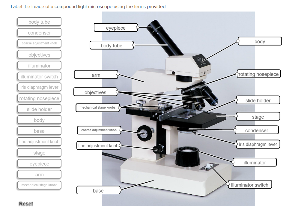
Solved Label the image of a compound light microscope using ...
Electron Microscopy Images - Dartmouth We have a library of images recorded using our scanning and transmission electron microscopes. Some are shown below and others elsewhere. These images are in the public domain. If you have questions about the images or want some specific images contact Max Guinel . Hibiscus Flower (August 2021) Morphy Amorphophallus titanum anther cross section.

Microscope Diagram Labeled, Unlabeled and Blank | Parts of a ...
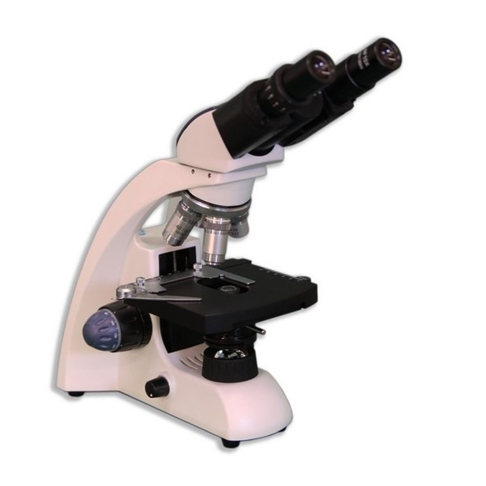
Parts of a microscope with functions and labeled diagram - Microbe Notes Parts of a microscope with functions and labeled diagram September 17, 2022 by Faith Mokobi Having been constructed in the 16th Century, Microscopes have revolutionalized science with their ability to magnify small objects such as microbial cells, producing images with definitive structures that are identifiable and characterizable.
Microscope With Labels - Free Transparent PNG Download - PNGkey
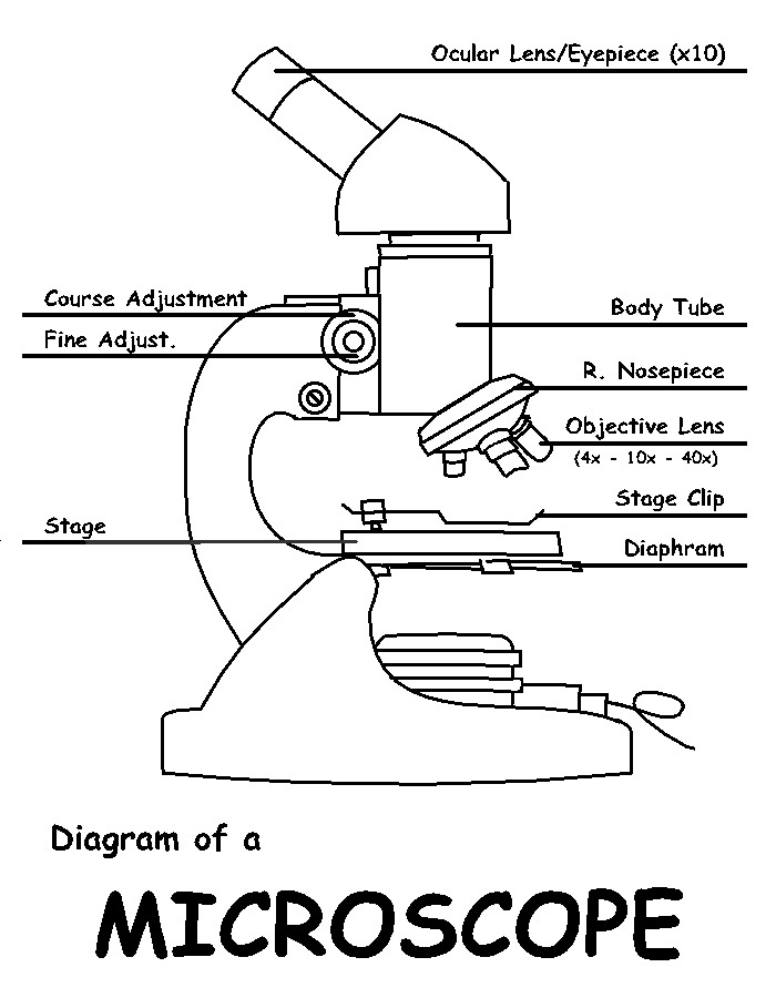
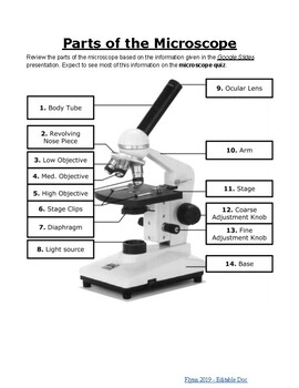
Parts of the Microscope with Labeling (also Free Printouts) Microscopes are specially created to magnify the image of the subject being studied. This exercise is created to be used in homes and schools. the microscope layout, including the blank and answered versions are available as pdf downloads. Click to Download : Label the Parts of the Microscope (A4) PDF print version.
Lab - Microscope: MAH-Summer 2019-Anatomy and Physiology I
PDF Label parts of the Microscope Label parts of the Microscope: . Created Date: 20150715115425Z

Label the parts of the microscope shown in the picture below ...
Lifestyle | Daily Life | News | The Sydney Morning Herald The latest Lifestyle | Daily Life news, tips, opinion and advice from The Sydney Morning Herald covering life and relationships, beauty, fashion, health & wellbeing
Microscope with labels picture
Two-photon excitation microscopy - Wikipedia Two-photon excitation microscopy (TPEF or 2PEF) is a fluorescence imaging technique that allows imaging of living tissue up to about one millimeter in thickness, with 0.64 μm lateral and 3.35 μm axial spatial resolution. Unlike traditional fluorescence microscopy, in which the excitation wavelength is shorter than the emission wavelength, two-photon excitation requires …
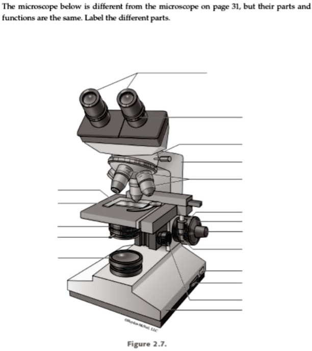
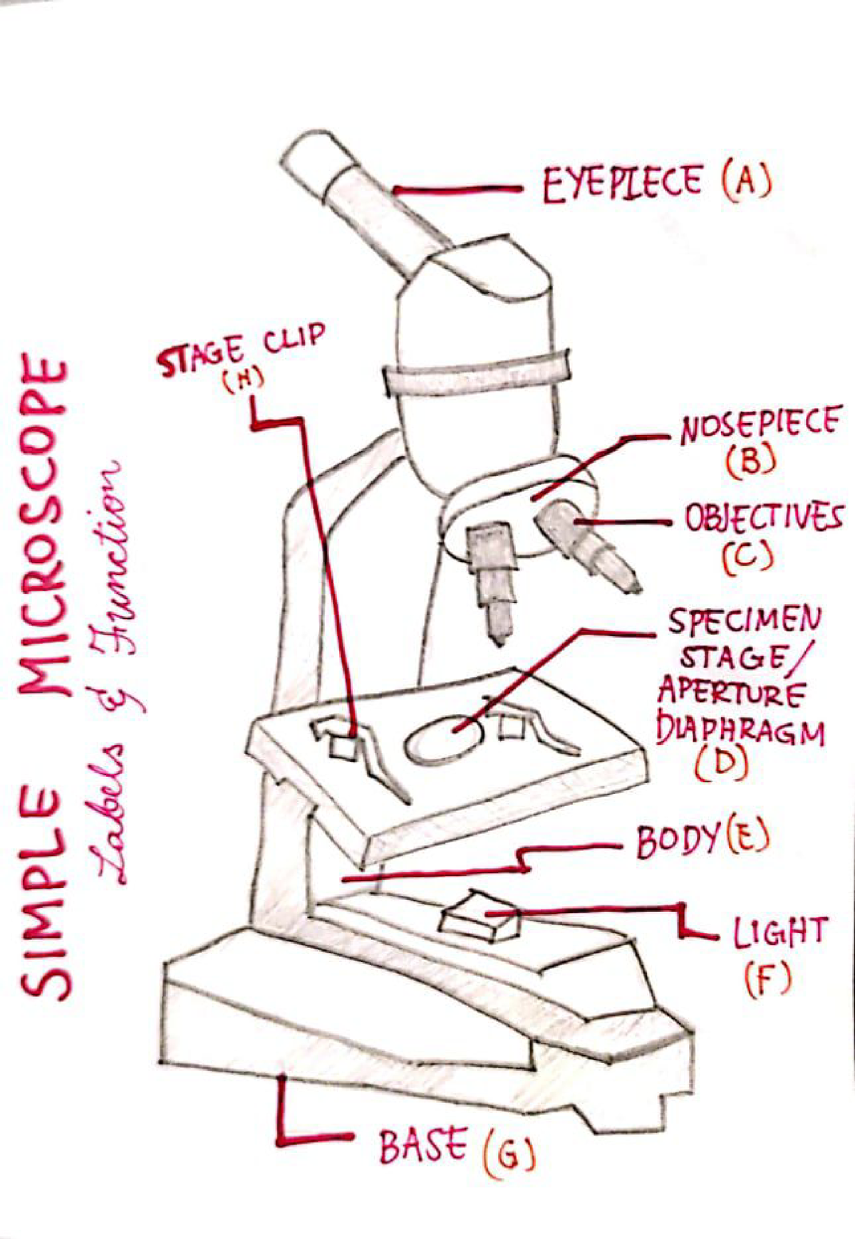
the microscope below i different from the micoscope on page 31 but their parts and functions are the sme label the different parts figure 27 93814
Technology and Science News - ABC News 7/12/2022 · The telescope was launched in December 2021 and sent back the first full-color images from its orbit 1 million miles from Earth. July 12 Pig organ transplants inch closer with testing in the dead
Free Microscope Drawing, Download Free Microscope Drawing png ...
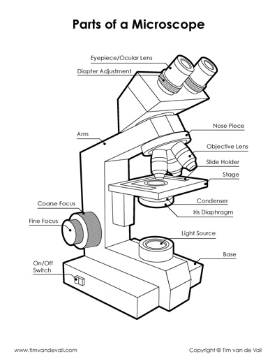
Microscope Parts, Function, & Labeled Diagram - slidingmotion Microscope parts labeled diagram gives us all the information about its parts and their position in the microscope. Microscope Parts Labeled Diagram The principle of the Microscope gives you an exact reason to use it. It works on the 3 principles. Magnification Resolving Power Numerical Aperture. Parts of Microscope Head Base Arm Eyepiece Lens

National Ecoline D-ELDB Binocular Digital Microscope
Scanning electron microscope - Wikipedia A scanning electron microscope (SEM) is a type of electron microscope that produces images of a sample by scanning the surface with a focused beam of electrons.The electrons interact with atoms in the sample, producing various signals that contain information about the surface topography and composition of the sample. The electron beam is scanned in a raster scan …
Label a microscope - Teaching resources
ZEISS Axioscan 7 Microscope Slide Scanner Digitize your specimens with Axioscan 7 – the reliable, reproducible way to create high-quality virtual microscope slides. Axioscan 7 combines qualities that you would not expect to get in a slide scanner: high speed digitization and outstanding image quality plus an unrivaled variety of imaging modes are all available in a fully automated and easy to operate system.
Microscope Diagram – Tim's Printables
ZEISS LSM 980 with Airyscan 2 – Confocal Microscope with … Specific areas of interest were imaged with the Airyscan 2 detector in order to acquire high resolution images of the Purkinje cells. The Airyscan 2 datasets were processed and orthogonal projections were created with ZEN Blue. The individual superresolution images were aligned with the cerebellum using ZEN Connect.

Biology Lab Microscope Labeling Diagram | Quizlet
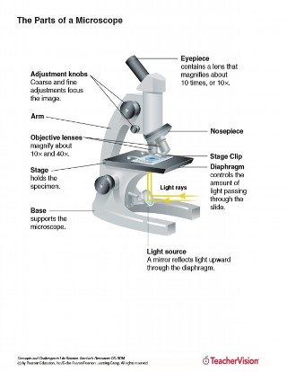
Compound Microscope Parts - Labeled Diagram and their Functions The eyepiece (or ocular lens) is the lens part at the top of a microscope that the viewer looks through. The standard eyepiece has a magnification of 10x. You may exchange with an optional eyepiece ranging from 5x - 30x. [In this figure] The structure inside an eyepiece. The current design of the eyepiece is no longer a single convex lens.

Microscope Labeling Diagram | Quizlet
Labeling the Parts of the Microscope | Microscope World Resources Labeling the Parts of the Microscope This activity has been designed for use in homes and schools. Each microscope layout (both blank and the version with answers) are available as PDF downloads. You can view a more in-depth review of each part of the microscope here. Download the Label the Parts of the Microscope PDF printable version here.

Dissecting Stereo Microscope Parts and Functions
Respiratory Histology Labeled | Virtual Anatomy Lab VAL - ncccval Body cavities, planes, and regions. Body Images Labeled. Body Images Unlabeled. Histology. Epithelium Images Labeled. Epithelium Images Unlabeled. Connective Tissue Images Labeled. Connective Tissue Images Unlabeled. Microscope.
The Parts of a Microscope (Labeled) Printable Printable (6th ...
Flow cytometry - Wikipedia Flow cytometry (FC) is a technique used to detect and measure physical and chemical characteristics of a population of cells or particles.. In this process, a sample containing cells or particles is suspended in a fluid and injected into the flow cytometer instrument. The sample is focused to ideally flow one cell at a time through a laser beam, where the light scattered is …

Microscope Labeling Practice Diagram | Quizlet

Meiji MT6500 Series PCM NIOSH 7400 Asbestos Microscope

Parts of Stereo Microscope (Dissecting microscope) – labeled ...
Microscope- Simple-AND Compound-WITH- Label - BS in Education ...

Lable the microscope worksheet

Types of Microscopes: Definition, Working Principle, Diagram ...

Parts of the Microscope with Labeling (also Free Printouts ...

Label Microscope Diagram - EnchantedLearning.com

Microscope - Label - Part 2 Diagram | Quizlet
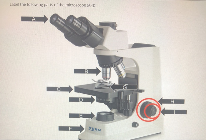
Solved Label the following parts of the microscope (A-1 ...

Biology Lab Quiz #2 (Labeling a Microscope) Diagram | Quizlet
Parts of the Microscope Labeling Activity!

A Study of the Microscope and its Functions With a Labeled ...

Microscope Labeling | PDF
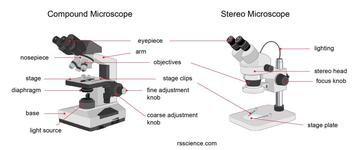
Parts of Stereo Microscope (Dissecting microscope) – labeled ...

microscope drawing with label - Clip Art Library
Compound Microscope Parts, Functions, and Labeled Diagram ...
Lab - Microscope: MAH-Summer 2019-Anatomy and Physiology I

Label the microscope — Science Learning Hub

Microscope With Labels free vector | Download it now!
Microscope labeling.pdf - ocular lens body tube revolving ...

Microscope With Labels clip art | Microscope parts ...

Below is a photo of a compound light microscope with labels ...

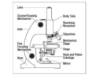
The Microscope

FUNRUI Kids Microscope, 450x, 200x, 100x Magnification Children Science Microscope Kit with LED Lights Includes Accessory Toy Set for Beginners Early ...
Post a Comment for "40 microscope images with labels"